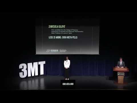
This research improves drug formulations by developing predictive tools for amorphous solid dispersions that increase drug solubility while allowing higher drug loading in a single tablet. The work aims to reduce pill burden, improve medication adherence, lower pharmaceutical development costs, and make treatments more effective for patients with chronic illnesses.
This research investigates how bacterial biofilms alter the mechanical properties of infected skin to improve microneedle-based drug delivery. By measuring tissue stiffness, structural integrity, and puncture resistance, it provides the evidence needed to design microneedles that can effectively penetrate biofilms, deliver antibiotics directly, and improve treatment of chronic wound infections.
2025
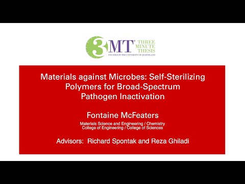
This research develops self-sterilising polymer coatings that become highly acidic when exposed to moisture, rapidly destroying harmful bacteria such as MRSA and E. coli. Designed for hospitals, classrooms, and other high-contact surfaces, these materials could reduce infections without harsh chemicals, helping prevent the spread of antibiotic-resistant bacteria.

This research challenges the long-standing assumption that brain regions causing no errors during awake brain surgery are functionally unimportant. By measuring subtle delays in speech rather than errors alone, it introduces causal parametric mapping, offering surgeons a more sensitive way to preserve language function and improve patient outcomes.
2025
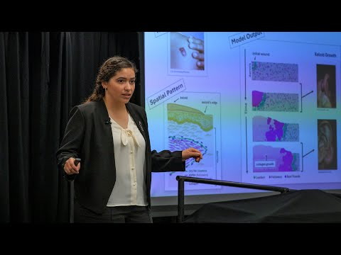
This research uses agent-based mathematical modelling to study keloid scar growth. By simulating interactions among collagen, immune cells, and key scar-associated cell types, the model predicts how keloids expand without requiring harmful patient experiments. The approach may guide future treatments for keloids and broader skin-healing conditions.

This research develops orally administered nanoparticle therapies for metronomic chemotherapy in ovarian cancer. By delivering smaller drug doses directly to tumours over extended periods, it aims to reduce side effects, overcome drug resistance, improve patient quality of life, and make long-term cancer treatment easier and more effective.

This research investigates whether the diabetes drug dapagliflozin (DAPA) can be repurposed to treat metabolic dysfunction-associated steatotic liver disease (MASLD). Using laboratory models, it examines fat accumulation and NHE1 ion channel function, aiming to develop a cost-effective treatment for two closely linked metabolic diseases with one existing medicine.
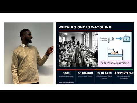
This research develops an ultra-low-power, battery-free newborn monitoring system for under-resourced hospitals. Using on-device artificial intelligence and energy harvesting, it continuously detects signs of distress while protecting patient privacy. The technology aims to support overstretched nurses, enable earlier intervention, and reduce preventable newborn deaths worldwide.

This research develops innovative three-dimensional "daisy" particle structures to improve inhaled medicines. Using Isothermal Dry Particle Coating, it prevents fine drug particles from clumping, ensuring they reach the lungs effectively. The work aims to improve inhaler performance and treatment for the 300 million people worldwide living with respiratory diseases.
2026
This thesis examines who turns to AI for mental health support, rather than whether AI can be a therapist. Drawing on TherapyGPT forum analysis and ongoing experiments, the research identifies fear of judgment, trust in AI and past therapist failures as possible drivers of AI therapy use.