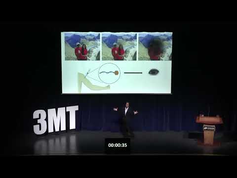
This research develops a targeted anti-VEGF therapy for wet age-related macular degeneration that can be injected under the skin rather than directly into the eye. In animal studies, the drug successfully reached the eye and reduced abnormal blood vessel growth, offering a safer, cheaper, and more convenient treatment for preventing blindness.

This research improves drug formulations by developing predictive tools for amorphous solid dispersions that increase drug solubility while allowing higher drug loading in a single tablet. The work aims to reduce pill burden, improve medication adherence, lower pharmaceutical development costs, and make treatments more effective for patients with chronic illnesses.
This research seeks blood-based biomarkers that predict which people infected with Chagas disease will later develop life-threatening cardiomyopathy. By analysing immune proteins in blood samples from Bolivia, it aims to enable earlier diagnosis, targeted monitoring, and preventative treatment, offering a model for predicting and preventing many chronic diseases before irreversible damage occurs.
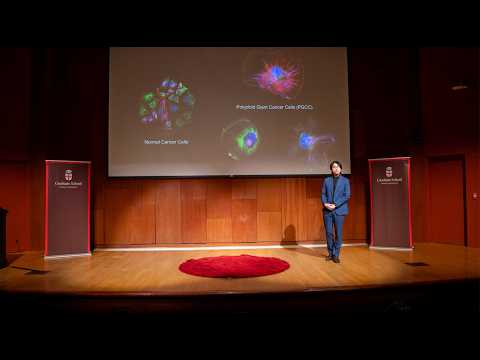
This research investigates polyploid giant cancer cells, a highly treatment-resistant population responsible for cancer relapse. By studying their structural biology and dependence on lipid metabolism, the work identifies metabolic vulnerabilities that can be targeted alongside chemotherapy, offering a promising strategy to eliminate resistant cancer cells and improve long-term treatment outcomes.
This research investigates a new targeted treatment strategy for kidney cancer by inhibiting the cancer-promoting protein PIM1 while enhancing TRAIL-mediated apoptosis. Together with the FDA-approved drug ONC201, this combination restores cancer cells' ability to self-destruct, offering a promising therapeutic approach now being evaluated in preclinical studies.
This research explores how early-life stress alters the gut microbiome and its communication with the brain, challenging the traditional "leaky gut" theory of anxiety. Using a comprehensive, lifespan-wide approach, it identifies a potential new mechanism that could enable more personalized treatments for patients who do not respond to current anxiety therapies.

This research identifies UCH-L1 as a promising blood biomarker directly linked to human eggs, offering a new way to measure ovarian reserve. A simple blood test could improve fertility assessment, detect premature ovarian aging earlier, and provide valuable insights into broader aspects of women's health, including cardiovascular health, cognitive aging, and longevity.

This research develops hybrid lipo-polymeric nanoparticles that overcome major limitations of current mRNA vaccine technology. The particles can be freeze-dried, rapidly loaded with mRNA, and simultaneously deliver therapeutic drugs. Their flexibility improves vaccine storage and distribution while enabling powerful combination therapies, including enhanced cancer treatments with improved survival in preclinical models.

This research develops an affordable, scalable platform for recording electrical activity from brain organoids. Using innovative basket-shaped sensors made from a low-cost conductive material, the system enables simultaneous recording from dozens of mini-brains, accelerating drug discovery and improving our understanding of brain diseases with more human-relevant laboratory models.

This research engineers DNA-modified exosomes to deliver drugs precisely to cancer cells while avoiding healthy tissue. By disguising natural cell-targeting signals and adding programmable DNA targeting molecules, the platform could reduce treatment side effects and provide a modular delivery system adaptable to many cancers and other diseases.