
This research develops a brain-inspired optical imaging system that mimics human vision to reconstruct objects hidden by fog, smoke, and biological tissue. Combining event-based cameras, spiking neural networks, and neuromorphic processors, it enables fast, energy-efficient imaging with applications in autonomous vehicles, emergency response, and non-invasive medical diagnostics.
This research develops advanced optical imaging technology to observe neurons firing in real time throughout the brain. By combining high-speed microscopy with flexible fibre-optic image relays, the system overcomes the challenge of light scattering, enabling clearer recordings of neural activity and deeper insights into brain function.

2026
This research investigates how Type 1 diabetes affects bone development during childhood and adolescence. Using high-resolution bone imaging and blood glucose data, the study explores whether blood sugar levels, variability, and disease duration influence bone health. Early findings suggest that diagnosis closer to puberty may be associated with lower bone density.
2026
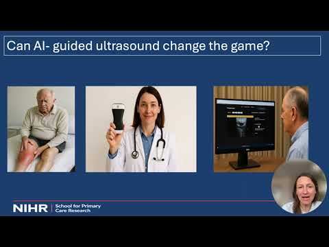
This research investigated whether AI-guided handheld ultrasound can help diagnose deep vein thrombosis (DVT) in primary care. Through a systematic review, a clinical study involving 565 patients, and stakeholder interviews, the research found promising results but highlighted challenges involving image quality, accountability, and integration into NHS healthcare systems.

This research develops a physics-based method for measuring lung elasticity from medical imaging to predict which emphysema patients will benefit from lung valve treatment. By creating detailed elasticity maps, the work aims to improve treatment selection, enhance patient outcomes, and provide new quantitative tools for assessing lung health.

This research develops nanobubble-enhanced ultrasound imaging as an accessible alternative to MRI for cancer diagnosis. Tiny gas-filled nanoparticles amplify ultrasound signals and improve image quality, particularly in prostate cancer. The technology could reduce diagnostic delays, lower costs, and provide high-quality medical imaging to more patients worldwide.

2026
This 3MT® presentation describes how artificial intelligence can help non-specialist clinicians diagnose deep vein thrombosis using AI-guided handheld ultrasound devices. By enabling faster point-of-care diagnosis in GP surgeries, the project aims to reduce hospital referrals, improve accessibility for vulnerable patients, and help healthcare systems manage increasing clinical demand more efficiently.

2025
This research uses artificial intelligence to predict the progression of Alzheimer’s disease and cancer using medical imaging data. By analyzing brain scans, tumor scans, and treatment responses, AI models can forecast disease development and treatment outcomes, enabling earlier intervention, more personalized care, and improved quality of life for aging populations.
2026
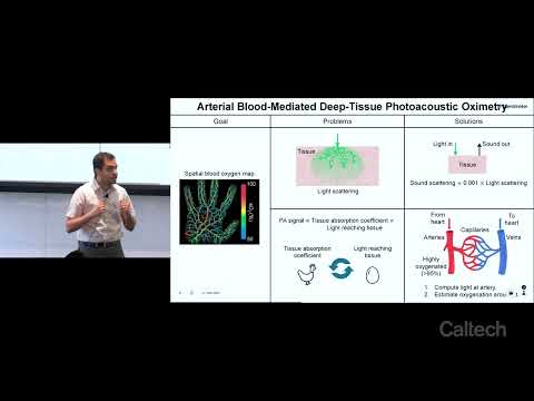
This research improves photoacoustic imaging, a technique that uses light-generated sound waves to visualize tissue oxygenation deep inside the body. By calibrating measurements using highly oxygenated arterial blood, the method overcomes longstanding accuracy limitations and avoids skin-tone bias, potentially improving early tumor detection and non-invasive monitoring of tissue health.

This research explores asthma by recreating lung airways using 3D bioprinting. By simulating low-oxygen conditions and imaging structural changes, it investigates how exaggerated immune responses narrow airways. These models enable detailed study of disease mechanisms and offer a platform to develop treatments, ultimately advancing efforts toward preventing or curing asthma.