
This research investigates how loneliness affects brain function across adulthood. Using brain imaging, it identifies age-related differences in activity within the caudate, a region involved in social reward processing. The findings suggest loneliness alters how people perceive social interactions, supporting the development of personalized, age-appropriate interventions to reduce chronic loneliness.

This research challenges the long-standing assumption that brain regions causing no errors during awake brain surgery are functionally unimportant. By measuring subtle delays in speech rather than errors alone, it introduces causal parametric mapping, offering surgeons a more sensitive way to preserve language function and improve patient outcomes.
2025
This research has developed a five-minute smartphone memory test that detects subtle cognitive changes associated with early Alzheimer's disease. The tool identified symptom-free individuals with underlying disease and predicted future cognitive decline, outperforming expensive brain scans while offering a simple, accessible, and affordable approach to early diagnosis.

2025
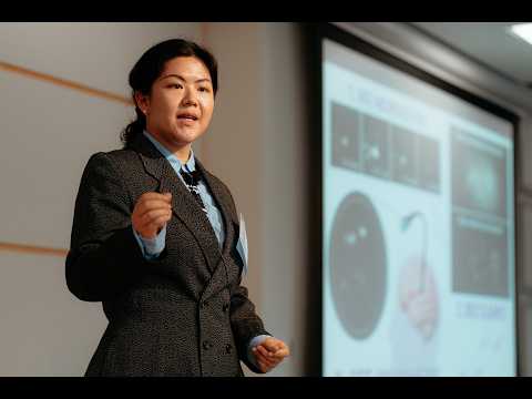
This research developed NanoX, a nanoscale fluorescent sensor that images oxytocin release from individual neurons in real time. By revealing patterns of brain chemistry associated with mental health disorders, the technology could enable earlier diagnosis, improve understanding of neurochemical signaling, and support both preventive and personalized mental healthcare.
This research develops advanced optical imaging technology to observe neurons firing in real time throughout the brain. By combining high-speed microscopy with flexible fibre-optic image relays, the system overcomes the challenge of light scattering, enabling clearer recordings of neural activity and deeper insights into brain function.
2025
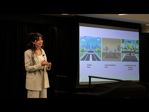
This research develops automated tools to identify psychedelic-inspired compounds that restore lost neural connections associated with depression, anxiety, and addiction. Using advanced imaging and custom analysis software, the project screens potential therapeutics that promote neuronal growth, aiming to create treatments that repair brain circuitry rather than simply managing symptoms.

2025
This research uses artificial intelligence to predict the progression of Alzheimer’s disease and cancer using medical imaging data. By analyzing brain scans, tumor scans, and treatment responses, AI models can forecast disease development and treatment outcomes, enabling earlier intervention, more personalized care, and improved quality of life for aging populations.

Tiny errors in electrode placement can determine success or failure of Parkinson’s surgery. This research develops high-resolution Polarization Sensitive Optical Tomography to map brain anatomy at micrometer scale—over 100 times finer than MRI. Automated scanning and 3D reconstruction create detailed connectivity maps, improving surgical precision and neuroscience understanding.
This research explores motor imagery as a rehabilitation tool after stroke. Brain imaging revealed sex-based differences in neural activation, with females showing greater efficiency. Practice improves patterns in both sexes. Understanding these differences enables personalized, home-based rehabilitation that may enhance recovery of arm and hand function.

My research explores whether people with semantic dementia can relearn everyday words through simple, repeated online training. Patients practiced picture–word pairs daily for two months and showed strong, lasting improvements that transferred to real-life use. The findings offer hope for patients and reveal how targeted practice can reshape the brain despite disease.